Depression Patients' Brain 'Salience Network' Is 73% Larger on Average, Seen in Childhood Before Onset
Individualized precision brain mapping showed that the frontostriatal salience network was markedly expanded in people with depression compared with healthy people.

When brains are compared as group averages, people with depression have not shown clear differences from healthy people. Researchers at Weill Cornell Medicine in the United States got past this limit with precision functional mapping, which scans one person dozens of times. They reported in Nature that the frontostriatal salience network, which handles reward and attention, is spread widely in people with depression.
Network Borders Expand Into Neighboring Regions
In several independent samples with repeated-scan data, the salience network covered nearly twice as much of the cerebral cortex in most people with depression, and this result was replicated across samples. The expansion mostly occurred as the network's borders pushed into neighboring regions, and it fell into three patterns from person to person.
This feature stayed stable over time regardless of mood state. In data from the U.S. Adolescent Brain Cognitive Development (ABCD) study, children who developed depression in adolescence already showed the expansion before onset. In a longitudinal analysis with up to 62 scans over a year and a half, changes in frontostriatal circuit connectivity moved along with symptom fluctuations and predicted later anhedonia.
Significance and Limits
The study points to a structural brain landscape tied to depression risk. However, because it is an observational study, it cannot tell whether the expansion causes depression, and the precision-scan samples are small, so more validation is needed before it can be used as a diagnostic tool.
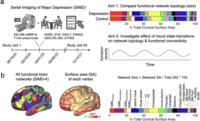
- Individualized precision functional mapping applied across several independent samples
- Salience network cortical area nearly doubled, stable regardless of mood state
- Expansion also seen in children before onset; circuit connectivity tracked symptom changes
Limitations This observational study does not establish causation, and the repeated-scan samples are too small for use as an individual diagnostic biomarker.
Original Research
- Lynch CJ, et al. · Weill Cornell Medicine. Frontostriatal salience network expansion in individuals in depression. Nature. DOI: 10.1038/s41586-024-07805-2
Copyright in the original research belongs to its publisher and authors.